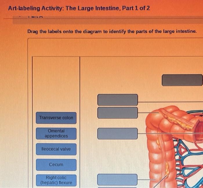
Art-labeling activity the gross anatomy of the large intestine – Embark on an art-labeling activity that unveils the gross anatomy of the large intestine, a crucial component of the digestive system. This interactive experience provides an immersive understanding of its structure, function, and clinical significance, fostering a deeper appreciation for the intricacies of human anatomy.
Delve into the external and internal features of the large intestine, identifying its segments and their distinguishing characteristics. Discover the arterial supply, venous drainage, nerve supply, and lymphatic drainage that sustain its function.
Gross Anatomy of the Large Intestine: Art-labeling Activity The Gross Anatomy Of The Large Intestine

The large intestine, also known as the colon, is the final portion of the digestive system. It plays a crucial role in the absorption of water and electrolytes, the formation and storage of feces, and the elimination of waste products from the body.The
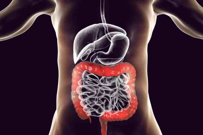
large intestine is approximately 5-6 feet long and extends from the ileocecal valve, where it connects to the small intestine, to the anus. It is located in the abdominal and pelvic cavities and is closely associated with other organs such as the stomach, liver, pancreas, and small intestine.
External Features of the Large Intestine
The large intestine has a characteristic appearance that distinguishes it from other parts of the digestive system. It is wider and shorter than the small intestine, with a diameter of about 2.5-7.5 cm. The surface of the large intestine is smooth and lined with numerous folds called haustra.
These haustra give the colon a sacculated appearance.The large intestine is divided into four main segments: the cecum, ascending colon, transverse colon, and descending colon. Each segment has distinct anatomical features and functions.
Internal Structure of the Large Intestine, Art-labeling activity the gross anatomy of the large intestine
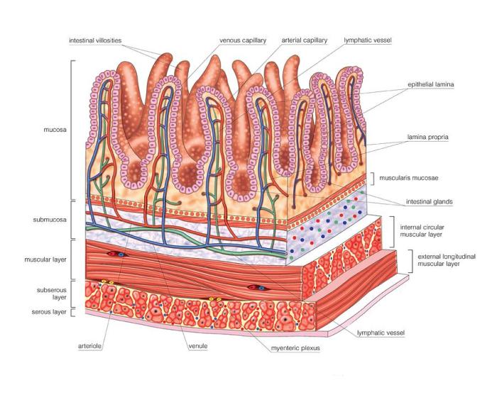
The internal structure of the large intestine is similar to that of the small intestine, consisting of four layers: the mucosa, submucosa, muscularis externa, and serosa. The mucosa is the innermost layer and is lined with a simple columnar epithelium.
The submucosa contains blood vessels, nerves, and lymphatic vessels. The muscularis externa is composed of two layers of smooth muscle that contract to propel feces through the colon. The serosa is the outermost layer and is composed of a thin layer of connective tissue that covers the colon and attaches it to surrounding structures.
Segments of the Large Intestine
The large intestine is divided into five segments:
- Cecum: The cecum is a pouch-like structure that is located at the junction of the small intestine and the large intestine. It is the first part of the large intestine to receive chyme from the small intestine.
- Ascending colon: The ascending colon extends from the cecum to the right side of the abdomen. It is about 15 cm long and ascends vertically.
- Transverse colon: The transverse colon extends from the ascending colon to the left side of the abdomen. It is about 50 cm long and crosses the abdomen horizontally.
- Descending colon: The descending colon extends from the transverse colon to the left side of the abdomen. It is about 25 cm long and descends vertically.
- Sigmoid colon: The sigmoid colon is the final segment of the large intestine. It is about 40 cm long and is located in the pelvic cavity. The sigmoid colon connects to the rectum, which leads to the anus.
Blood Supply and Innervation of the Large Intestine
The large intestine is supplied with blood by the superior mesenteric artery and the inferior mesenteric artery. The superior mesenteric artery supplies the cecum, ascending colon, and transverse colon. The inferior mesenteric artery supplies the descending colon, sigmoid colon, and rectum.The
large intestine is innervated by the autonomic nervous system. The parasympathetic nerves supply the large intestine with motor innervation, which controls the movement of feces through the colon. The sympathetic nerves supply the large intestine with sensory innervation, which transmits pain and other sensations to the brain.
Clinical Significance of Large Intestine Anatomy
Understanding the anatomy of the large intestine is important for several reasons:
- Diagnosis and treatment of diseases: Knowledge of the large intestine anatomy is essential for diagnosing and treating diseases that affect the colon, such as colorectal cancer, diverticulitis, and inflammatory bowel disease.
- Surgical procedures: Surgeons need to have a thorough understanding of the large intestine anatomy in order to perform surgical procedures such as colonoscopy, colectomy, and colostomy.
- Imaging studies: Radiologists use knowledge of the large intestine anatomy to interpret imaging studies such as X-rays, CT scans, and MRIs.
FAQ Corner
What is the purpose of the large intestine?
The large intestine absorbs water and electrolytes from undigested food, forming feces and preparing them for elimination.
What are the main segments of the large intestine?
The main segments of the large intestine are the cecum, ascending colon, transverse colon, descending colon, and sigmoid colon.
What is the clinical significance of understanding the anatomy of the large intestine?
Understanding the anatomy of the large intestine is essential for diagnosing and treating conditions such as appendicitis, diverticulitis, and colon cancer.


